
What does a Mesozoic dinosaur unearthed in Kansas have in common with John the Baptist and hundreds of millions of people worldwide have in common? Osteoarthritis. Not to be confused with its sister diseases, rheumatoid arthritis (inflammation caused by autoimmune reaction in the joints) and osteoporosis (the gradual weakening of bones), osteoarthritis (OA) is the gradual thinning of the articular cartilage — the smooth, lubricating connective tissue that covers the ends of bones at a joint. Articular cartilage provides pain-free movement at the joints, at least when it’s healthy.
When cartilage becomes damaged and thin, bond grinds against bone. In some patients, you can actually hear a crackling noise as the bones grate against each other. Over time, the friction wears down the bone until nerve ends are exposed, and everyday movements — walking, sitting, even writing — result in excruciating pain.
In the latest stages of OA, after all other options for pain relief have failed, the last resort is joint replacement. This has three major problems: First, joint replacement doesn’t significantly reduce the pain for 10% of patients. Second, 10% of joint replacements fail within 15 years and need to be replaced. And third, about 1% of patients develop an infection due to the surgery. While these percentages may seem relatively small, there are over 1 million joint replacement surgeries in the U.S. alone every year. This means hundreds of thousands of people either have complications associated with joint replacement or don’t experience pain relief.
The medical community has worked for over a century to develop strategies for repairing the damaged cartilage before surgery is needed. However, these strategies have significant flaws — most notably, short-term repair and long recovery periods. According to a recent study in npj Regenerative Medicine, researchers have developed a strategy without these pitfalls — a stem cell-based bioimplant.
We can repair cartilage by drilling holes in bones, but should we?
The traditional explanation of OA-associated articular cartilage damage was “mechanical trauma” — the unavoidable “wear and tear” of aging and overuse. However, over the last several decades our understanding has shifted; we now know that OA is multifactorial in origin. Mechanical trauma does play a role, but so do genetics, nutrition, diabetes, immunity, limb alignment, and even joint shape. Regardless of the underlying mechanism, one aspect remains the same: OA implies articular cartilage damage.
The body is not great at repairing articular cartilage. Prior to the 1950s, there was no established medical technique that provided much help. Patients either endured the pain or underwent risky joint replacement surgery (with dubious results). However, that changed in 1959, when a seven-sentence article appeared in the Proceedings of the Congress of the British Orthopaedic Association. In the surprisingly concise article, Dr. Kenneth H. Pridie, a British clinician who was treating OA patients, noted that “If [ends of bones] were drilled and the holes were not too far apart, smooth [cartilage] would spread over the surface.”
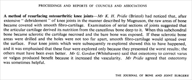
Shortly after publishing this revolutionary procedure, Pridie died, leaving scientists to ponder where he came up with this idea and who would allow him to perform this operation, as well as more practical questions, such as “how big of a drill bit?” and “how far apart is too far apart?”
Six decades later, scientists are still working on answering these questions. However, researchers in the 1980s uncovered the basic mechanism underlying Pridie’s technique: By drilling through the ends of the bone, cells in the bone marrow could access the damaged joint and generate new cartilage. Currently, this technique — called microfracture — is the most common treatment for regenerating articular cartilage. It’s a quick procedure (typically lasting between 30 to 90 minutes), minimally invasive, and the recovery time is relatively short (four to seven months).
But there is a downside: This new cartilage is biomechanically inferior to the original articular cartilage. As a result, it can wear out after just a few years, and the procedure needs to be repeated, which increases the likelihood of a complication (such as infection). Denis Evseenko — a professor of Orthopedic Surgery, Stem Cell Research, and Regenerative Medicine at Keck School of Medicine of USC — wanted his bioimplant to create strong articular cartilage that could last for decades. To do this, he had to find a way to harness the power of the lone cell that can create articular cartilage.
Only one cell knows the recipe for articular cartilage
Cartilage is one of the simplest connective tissues of the body. Or, at least, it is simple in terms of its ingredients: water and a handful of different proteins (several of which you likely have in your pantry). But there is only one chef who can whip up articular cartilage just right: the chondrocytes.
Chondrocytes live a life of solitude. Embedded in the cartilage, each cell governs its own microenvironment where it alone is responsible for maintaining the cartilage in that region. We’re still learning about these cells, but we know that when they can no longer fulfill their role, there is rarely a replacement. While some scientists were working to understand how bone marrow cells could be used to regenerate cartilage, other scientists were looking into how to harness chondrocytes for the same purpose.
A new technique emerged in the 1980s: A surgeon removes a small segment of bone from a patient, extracts a few chondrocytes, and replicates them in a petri dish. After a sufficient number of chondrocytes are generated, the surgeon implants them back into the patient. The cartilage that results from this procedure is just as strong as the original. However, this technique has failed to gain much momentum because it requires multiple surgeries, and it can take up to two years for the implanted chondrocytes to repair the cartilage.
One of the reasons for the long recovery time is the use of “old” chondrocytes. As a person gets older, chondrocytes slow down their cartilage production. When an old chondrocyte replicates, it creates another chondrocyte that acts like an old chondrocyte (in that cartilage production is slow). Researchers had long suspected that chondrocytes created from stem cells would act like a young chondrocyte (with fast cartilage production), but no one knew how to create stem cell-derived chondrocytes until 2010.
If the body wants stem cells to create chondrocytes, it delivers carefully timed signals to the stem cell. Give the wrong signal at the wrong time, and you might have a totally different type of cell. Determining the correct signals and the correct timing is a tough problem to crack. But in 2010, a group of researchers at University of Manchester did just that: They created chondrocytes from stem cells.
Armed with this new knowledge, Evseenko and his group of researchers set out to design a therapeutic bioimplant that captures the best of the two current strategies for cartilage regeneration: a minimally invasive quick procedure with a short recovery time that produces strong articular cartilage. A tall order, but equipped with stem-cell derived chondrocytes, they succeeded in 2018.
Their bioimplant was composed of a cartilage-like matrix, embedded with stem cell-derived chondrocytes. Four weeks after being applied to a mouse knee joint, the chondrocytes had replaced their cartilage-like environment with strong articular cartilage. These results were promising, but the joints of small animals are structurally different from large animals, including humans. Most notably, small animals have very thin articular cartilage. So before they moved on to clinical trials in humans, the researchers needed to scale up to something larger than a mouse: the Yucatan minipig.
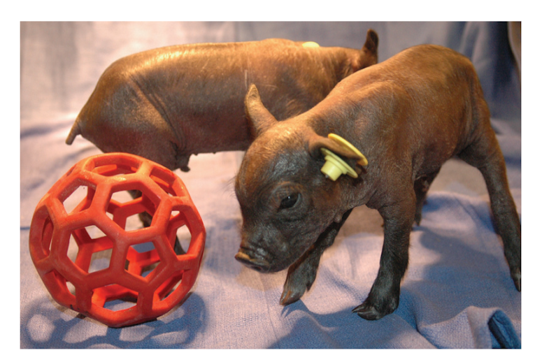
A “minipig” may not sound like a great example of a large animal, being only about 16 inches tall and 36 inches long. However, they weigh approximately 160 lbs. To cushion all that weight on their little joints, their articular cartilage is very thick, similar to that of humans.
To mimic OA-associated articular cartilage damage, the researchers cut away segments of articular cartilage at the knee joint. They applied their bioimplant to the area. The pigs recovered for six months, and when the researchers looked at the joints again, it was immediately clear that the articular cartilage regenerated in pigs treated with the implant. Most importantly, the cartilage was thick and shock absorbent, far superior to the cartilage produced by the microfracture technique.
The work will now advance into humans with support from a $6 million grant from the California Institute of Regenerative Medicine.